The mucosal defences triggered by S. Typhimurium in vivo
We have developed the "streptomycin mouse model" for Salmonella diarrhea (Barthel, Hapfelmeier et al., 2003; Kaiser et al., 2012). This robust experimental system has transformed the way we study the interplay of virulence factors and mucosal host responses in the complex scenario of a real infection. Most importantly, this animal model has provided a bridge to the field of mucosal immunology. Thereby, the research on Salmonella diarrhea can benefit from the highly advanced repertoire of genetically modified animals and analytic tools developed by immunologists.
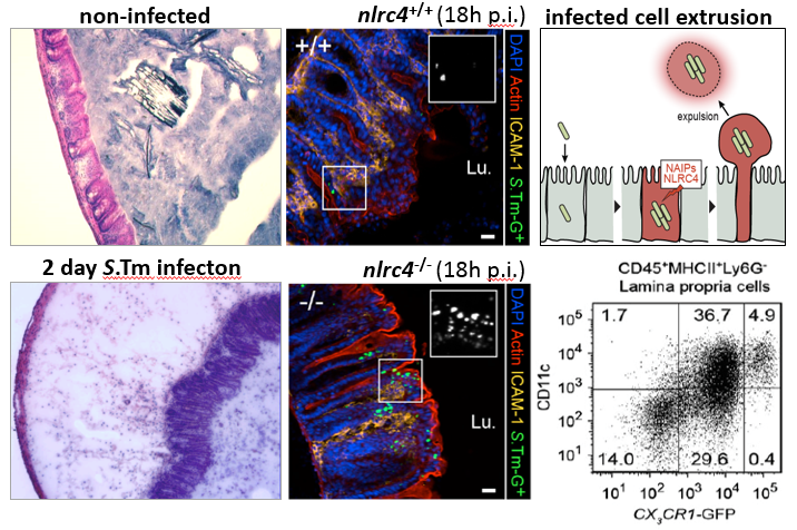
Using this model, we have determined the role of S. Typhimurium virulence factors and discovered a key role of mucosal dendritic cells (Hapfelmeier et al., 2008). The immunological approach is further complemented by powerful in vivo imaging techniques, e.g. intravital microscopy to follow the infection at the single cell level in real time within live tissues (Müller et al., 2012; Sellin et al., 2014). Recently, we discovered a central role of the so-called NAIP/NLRC4/caspase-1 inflammasome, in protecting the gut epithelium from infection (Sellin et al., 2014; Sellin et al., 2015). Based on these findings, we are now probing the molecular cascades and cellular events eliciting disease upon bacterial invasion of the epithelium. We anticipate that these results will have important implications for handling of commensals and pathogens by the mucosal innate immune system.
Combining knockout mouse models with state of the art light microscopy will be of great value for deciphering the disease-triggering cellular interactions between the pathogen and the host's mucosal immune system. Our goal is a new type of "in vivo cellular microbiology".
Intravital microscopy
Powerful in vivo imaging techniques allow us to follow the Salmonella infection at the single cell level in real time within live tissues (external pageMüller et al., 2012; external pageSellin et al., 2014).
Literature
Barthel, M., S. Hapfelmeier, L. Quintanilla-Martinez, M. Kremer, M. Rohde, M. Hogardt, K. Pfeffer, H. Russmann, and W. D. Hardt*. 2003. Pretreatment of mice with streptomycin provides a Salmonella enterica serovar Typhimurium colitis model that allows analysis of both pathogen and host. Infect Immun 71:2839-58. external page[Abstract]
Hapfelmeier, S., A. J. Müller, B. Stecher, P. Kaiser, M. Barthel, K. Endt, M. Eberhard, R. Robbiani, C. A. Jacobi, M. Heikenwalder, C. Kirschning, S. Jung, T. Stallmach, M. Kremer and W. D. Hardt*. 2008. Microbe sampling by mucosal dendritic cells is a discrete, MyD88-independent step in ΔinvG S. Typhimurium colitis. J Exp Med. 205(2):437-50. external page[Abstract]
Kaiser, P., Diard, M., Stecher, B. and W.D. Hardt* (2012) The streptomycin mouse model for Salmonella diarrhea: functional analysis of the microbiota, the pathogen’s virulence factors and the host’s mucosal immune response. Immunol. Rev., 245(1):56-83. external page[Abstract]
Müller, A.J., Kaiser, P., Dittmar, K.E.J., Weber, T.C., Haueter, S., Endt, K., Songhet, P., Zellweger, C., Kremer, M. , Fehling, H.J. and W.D. Hardt* (2012) Salmonella Gut Invasion Involves TTSS-2-Dependent Epithelial Traversal, Basolateral Exit, and Uptake by Epithelium-Sampling Lamina Propria Phagocytes. Cell Host Microbe 11(1):19-32. external page[Abstract]
Sellin, M.E., Müller, A.A., Felmy, B., Dolowschiak, T., Diard, M., Tardivel, A., Maslowski, K.M. and W.D. Hardt* (2014) Epithelium-intrinsic NAIP/NLRC4 inflammasome drives expulsion of infected enterocytes to restrict Salmonella replication in the intestinal mucosa. Cell Host Microbe, 16(2):237-48. external page[Abstract]
Sellin, M.E.*, Maslowski, K.M., Maloy, K.J. and W.D. Hardt* (2015) Inflammasomes of the intestinal epithelium. Trends Immunol. 36(8):442-50. external page[Abstract]
Dolowschiak T., Mueller A.A., Pisan L.J., Feigelman R., Felmy B., Sellin M.E., Namineni S., Nguyen B.D., Wotzka S.Y., Heikenwalder M., von Mering C., Mueller C., Hardt W.D. (2016) IFN-γ Hinders Recovery from Mucosal Inflammation during Antibiotic Therapy for Salmonella Gut Infection. Cell Host Microbe. 2016 Aug 10;20(2):238-49. external page[Abstract]
Sellin M.E., Müller A.A., Hardt W. D. (2017) Consequences of Epithelial Inflammasome Activation by Bacterial Pathogens. J Mol Biol. 2017 Apr 25. external page[Abstract]